Let’s continue with the macroscopic and histological characteristics of the most relevant lesions in porcine respiratory processes

Macroscopic and histological characteristics of the most relevant lesions in porcine respiratory processes

There are three most common forms of rhinitis:

It is generally associated with Porcine Cytomegalovirus. Sometimes, only congestion is seen, so the diagnosis is histological and is based on the observation of large viral inclusion bodies in the epithelial cells (“inclusion body rhinitis”).

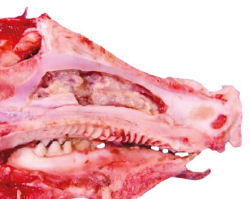
Image 5. Detail of suppurative exudate in a longitudinally open nasal cavity.


In the swine species, lesions in these anatomical areas are infrequent, although inflammatory or vascular lesions (petechial hemorrhages, edema) can be observed in systemic diseases (classical swine fever, salmonellosis, edema disease).


Melanosis, characterized by black dots or spots of few centimeters in diameter on the lung surface, is not clinically significant, but is a frequent finding in slaughterhouses. In this case, the texture of the pigmented lungs remains unchanged.

Atelectasis, or incomplete distension of the alveoli and lack of lung expansion, may be distributional:
Diffuse (congenital or neonatal atelectasis): is due to lack of expansion of the lungs with air at the time of birth.
Focal or locally extensive: this is usually an acquired condition, in which the lungs collapsed after inflation due to obstructive or compressive causes.
Generally, the affected areas become dark red, firm, “fleshy” (due to lack of air) and moist (fetal fluid).
Atelectasis and pneumonia have a similar macroscopic appearance. In many cases, tissue with atelectasis is depressed with respect to the adjacent healthy tissue, while with pneumonia the affected tissue is similarly or elevated with respect to the unaffected tissue.
Pulmonary emphysema is the permanent widening or dilation of the lung due to increased air content. In pigs, it is more common in the interlobulillary (interstitial) septa due to violent respiratory efforts (terminal or agonal emphysema).

Hemorrhages, thrombosis and infarction are associated with trauma, coagulopathies, disseminated intravascular coagulation (DIC), vasculitis, sepsis and pulmonary thromboembolism.
Pulmonary edema is characterized by the accumulation of fluid in the alveoli (alveolar edema) or in the pulmonary interstitium (interstitial edema).
Macroscopically, the lungs appear to be moist and heavy, regardless of the cause, with increased lung volume/size. The color varies, depending on the degree of congestion or hemorrhage, with the presence of foamy fluid in the trachea and bronchi being common.

Inflammations or pneumonias are the most common processes in pig lungs. Based on texture, distribution, appearance and type of exudate, pneumonias can be roughly classified into four morphologically distinct types:
Bronchopneumonia
Interstitial Pneumonia
Embolic Pneumonia
Granulomatous Pneumonia
These four morphological types allow the clinician or pathologist to predict the most probable etiology, facilitating decision making regarding sampling and tests to be requested from the diagnostic laboratory (histopathology, bacteriology, virology or toxicology). However, it is possible for these four types of pneumonias to overlap, and sometimes two morphological types may be present in the same lung. The histological study can help in many of these cases.
The most relevant and differential morphological characteristics of each of them are shown in Figure 1 and Table 1.
Figure 1. Morphological patterns of pneumonia.
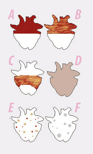
A) Catarrhal or suppurative bronchopneumonia
B) Fibrinous bronchopneumonia
C) Porcine pleuropneumonia
D) Interstitial pneumonia
E) Embolic pneumonia
F) Granulomatous pneumonia
Table 1. Types of pneumonia. Morphological and differential characteristics.
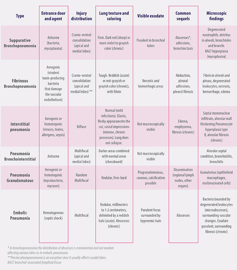
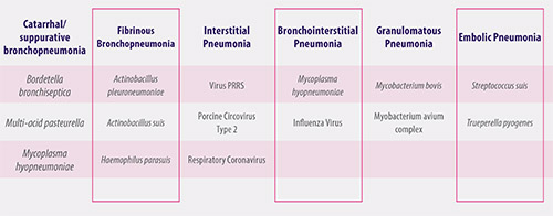
The pathogens most commonly implicated in each type of pneumonia are shown in Table 2. With this simple classification it is possible, at the time of the necropsy, to predict with some degree of certainty:
The probable cause: virus, bacteria, fungi or parasites
The entry route: aerogenic or haematogenic
The possible consequences
Table 2. Main etiological agents and morphological types of pneumonia.

In the case of bronchopneumonia, the inflammatory lesion is centered in the alveolus, and two different types can be found:
Suppurative or purulent bronchopneumonia, also called catarrhal or catarrhal-purulent in earlier phases (Image 6).
Fibrinous bronchopneumonia, also called pleuropneumonia (Image 7).
Image 6. Severe, extensive suppurative bronchopneumonia, chronic, with abscess formation and fibrous adhesions. Histologically, degenerated leukocytes and detritus occupy alveoli and bronchial spaces.
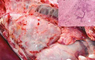
Image 7. Subacute, extensive fibrinous bronchopneumonia.

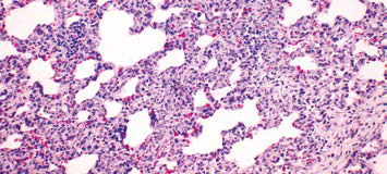
Of all the morphological types, it is the most difficult pneumonia to diagnose macroscopically. It is an inflammation of alveolar walls and lung interstitium, with or without macroscopic changes depending on the intensity or chronicity of the process. The histological study is especially important in these cases (Image 8).
Image 8. Histological image of interstitial pneumonia with thickening of alveolar septa by infiltration of mononuclear leukocytes.
It can also present with multifocal distribution, usually by alveolar and bronchial affection: bronchointerstitial pneumonia (Image 9). 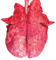
In many cases, virus-induced lesions can be masked by changes resulting from concurrent or secondary bacterial infections.
Figure 9. Bronchointerstitial pneumonia (chessboard) by Mycoplasma hyopneumoniae. The pneumonic pattern in cases of Swine Influenza is very similar and macroscopically indistinguishable.

In the case of embolic pneumonia, an inflammatory pattern is observed that is characteristic of bacterial septicemic processes that reach the lung parenchyma via hematogenous, producing multifocal lesions of random distribution (Image 10).
Image 10. Embolic Pneumonia. Detailed observation of one of the typical lesions, purulent focus surrounded by hyperemic, reddish halo.
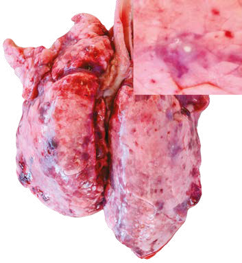

Granulomatous pneumonia is characterized by lung consolidation associated with the formation of granulomas, being related to elements of difficult elimination such as mycobacteria, fungi, parasites and foreign bodies.

The inflammatory condition of the pleura, without or with minimal superficial pulmonary involvement, is common in the porcine species and is usually part of disseminated processes such as polyserositis, polyarthritis and meningitis, being common in infections by Haemophilus parasuis, Streptococcus suis and Mycoplasma hyorrhinis.
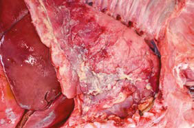
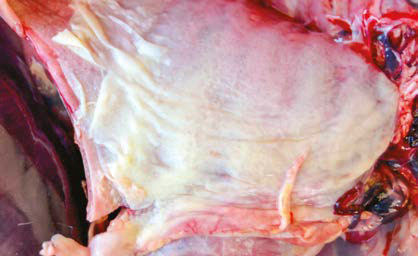
Macroscopically, it is characterized by extensive plaques and fibrin aggregates in the most acute or subacute forms (Image 11), which evolve over time to fibrous adhesions with the costal wall or pericardium.
Image 11. Fibrinous pleuritis in Glässer’s disease.
The keys to sampling for histopathology
The microscopic examination allows the pathologist to evaluate whether the agents isolated in the tissues are significant contributors to the lesion and also whether there is involvement of other agents not detected in the macroscopic examination.
The immunohistochemistry (IHC) is an additional tool to the microscopic study, very useful to detect some pathogens not visible or difficult to recognize microscopically, or when they are in low numbers, especially viral agents.
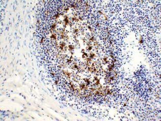
IHC also allows direct visualization of the location of the antigen in the tissue (Image 12).
Tissue sections are prepared in a similar way to that used for histopathology, but then stained with antiserum against a specific agent.
Due to the expense and labor involved in the technique, it should always be preceded by a histopathological study that evidence of injuries related to the agent to be identified.
Image 12. Immunohistochemistry. PCV2 antigen in mediastinal lymph node (in brown).
The main characteristic that a sample for histopathological analysis must have is its representativeness of the process to be assessed (taking from an appropriate area or another that provides more information) and the integrity of the tissue to be assessed (correct fixation).

For this, some important general considerations must be taken into account:
1.Preservation of the sample by adequate fixation
Autolytic tissue is not suitable for any histopathological test, which implies
Fixing the samples in formalin: commercial formalin (normally 35-40%) should be diluted in water at 1:10. Currently, it is common to commercialize ready-to-use formalin (4% concentration).
The samples in formalin do not require refrigeration, they are perfectly maintained at room temperature.
NEVER FREEZE! Routine freezing causes the formation of crystals in the tissue that damage it, creating serious artifacts that can make the sample unvaluable.
Respect a minimum amount of fixative in relation to the volume of sample in the container.
We must ensure that the proportion of tissue:formaldehyde is always from 1:5 to 1:10. If this proportion is not respected, the fixative will be insufficient and the tissue will suffer autolysis, making it unsuitable for histopathological evaluation.
2. Characteristics of the sample
Usually 3-5 samples are taken from different lobes of the lung, including injured and normal areas.
The most recommended way is to take from the transition areas between injury and healthy tissue, as these areas usually concentrate a greater number of viable pathogens and allow observation and study the type of reaction of the body to the pathogen or damage (types of exudates, inflammatory cells, fibrosis, reparative phenomena, etc.).
It is also advisable to take samples from the apical and medial lobes, usually where microscopic lesions are larger and evident.
The size of the samples should not exceed 1-2 cm in thickness, which is the penetration capacity of formaldehyde.
It is recommended to take sheets or slices of that thickness. If the samples are larger, the formaldehyde will not penetrate the central areas and will undergo autolysis.
Note that chronic and more acute lesions may be present in the same lung and may represent the activity of different pathogens.
3. Sending samples


Bibliographic references and further reading recommended are available upon request
Subscribe now to the technical pig magazine
AUTHORS
Bifet Gracia Farm & Nedap – Automated feeding in swine nurseries

The importance of Water on pig farms
Fernando Laguna Arán
Microbiota & Intestinal Barrier Integrity – Keys to Piglet Health
Alberto Morillo Alujas
Impact of Reducing Antibiotic use, the Dutch experience
Ron Bergevoet
The keys to successful Lactation in hyperprolific sows
Mercedes Sebastián Lafuente
Addressing the challenge of Management in Transition
Víctor Fernández Segundo
Dealing with the rise of Swine Dysentery
Roberto M. C. Guedes
Actinobacillus pleuropneumoniae – What are we dealing with?
Marcelo Gottschalk
The new era of Animal Welfare in Pig Production – Are we ready?
Antonio Velarde
Gut health in piglets – What can we do to measure and improve it?
Alberto Morillo Alujas
Interview with Cristina Massot – Animal Health in Europe after April 2021
Cristina Massot
Differential diagnosis of respiratory processes in pigs
Desirée Martín Jurado Gema Chacón Pérez